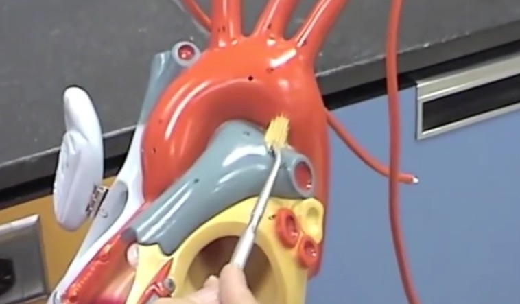
Also called the arterial ligament, the ligamentum arteriosum is a small ligament connect to the superior surface of the proximal descending aorta and the left pulmonary artery. It develops about 3 weeks after birth and is considered as a non-functional part of the ductus arteriosus.
The ligamentum arteriosum is related closely to a left vagus nerve branch referred to as the left recurrent laryngeal nerve. After the left recurrent laryngeal nerve splits from the left vagus nerve, it hooks around the aortic arch behind the ligamentum arteriosum, and then goes up towards the larynx.
The ligamentum arteriosum has a part to play in major injuries or trauma. It corrects the aorta and fixes it in place at the time of rapid decelerations recoil, subsequently probably causing the aorta to rupture.
Ligamentum arteriosum – Structure and Clinical significance
The ligamentum arteriosum is a bit of fibrous tissue which forms from the DA or ductus arteriosus. If develops about 3 weeks after birth and then becomes a part of the normal adult cardiac structure.
The arterial ligament is described by some as a non-functional piece of the DA after the formation of the embryo. Others however consider the ligament as one of the factors which contribute towards aorta rupture during times of major trauma. It is said that the ligamentum arteriosum keeps the largest artery in the body in its place when it comes back to its normal location at the time of rapid deceleration. This can sometimes cause the artery to burst open.
At the time of prenatal formation, the fetal heart features the ductus arteriosus, which is a small passage or a hole. The DA joins the aortic arch and the pulmonary artery which transfers de-oxygenated blood to the lungs from the heart. However, in a fetus, it performs the function of allowing the passage of a large quantity of blood from the right ventricle to around the lungs filled with fluids.
The DA starts closing in 12 to 24 hours after a person’s birth. This process normally finishes in about 3 weeks. After the process is complete, it forms into the ligamentum arteriosum, which is attached to the pulmonary artery and the arch of the aorta. The aortic arch is located postero-superior, i.e., above and to the back of the ligamentum arteriosum, while the pulmonary artery is position towards its lower middle region.
In case of a patent ductus, surgery would involve incision of the pleura above the aortic arch, back of the vagus nerve, and above near the starting point of the left sub-clavian artery. The vagus, the pleural flap, and the left recurrent laryngeal branch of the vagus are reflected forwards to permit adequate ductus access.
The shriveled remains of the DA, i.e., the ligamentum arteriosum, passes from the origins of the pulmonary artery to the aortic arch’s concave area, beyond the branching off point of the sub-clavian artery. It almost lies horizontally and the left recurrent laryngeal nerve loops around it. The shallow section of the heart plexus is located in front of it, while behind and deeper towards the right is the left main bronchus.
There are 12 paired cranial nerves and the tenth one is called the vagus nerve which is located lateral to the ligamentum arteriosum tissue. This is also why the vagus nerve is referred to as cranial nerve X. Galen’s nerve or the recurrent laryngeal nerve is also situated laterally; it springs from the left of the vagus nerve and loops around the ligamentum arteriosum from the back where a section of the aorta is present. Later it goes upwards towards the voice box from which it gets its name.
Towards the front, the ligament is located next to the surface of the cardiac plexus which is a tangle of nerves that play a part in innervating the heart from its basal area. The phrenic nerve is also located in the front section of the ligament; it typically springs from the C4 or the 4th cervical nerve.
Difference between ductus arteriosus and ligamentum arteriosum
There is no expansion of the lungs in the fetus. Hence, most blood from the cardiac right ventricle is passed to the aorta from the pulmonary artery via the patent ductus arteriosus. After birth, the lungs expand with just a few breaths and the blood passes to the pulmonary artery from the right ventricle into the lungs. Subsequently, the bradykinins released from the infant’s expanding lungs and withdrawal of the mother’s circulating prostaglandin causes the DA to close in some minutes or hours. Eventually, the DA develops into the ligamentum arteriosum ligament.
- It may be noted that even though the DA normally closes after birth and forms the ligamentum arteriosum, the closure may not occur in some people. Such patients are treated for PDA during infancy.


Накрутка Twitch Зрителей
Где Вы ищите свежие новости?
Лично я читаю и доверяю газете https://www.ukr.net/.
Это единственный источник свежих и независимых новостей.
Рекомендую и Вам