The hyoid bone refers to a bone located in the front middle section of the neck, between the thyroid cartilage and the chin. The name has its origins in the Greek word ‘hyoeides’ which means shaped like the Greek letter ‘υ’ (upsilon). It is shaped like a horseshoe and is one of the few bones in the human body that is not connected to any other bone. At rest, the hyoid bone lies posteriorly to the C3, i.e., the 3rd cervical vertebra, and anteriorly at the level of the bottom section of the mandible.
The hyoid bone, unlike other types of bones, is articulated only distantly via ligaments or muscles to other bones. It is held in place in the back, front, and bottom directions by varied muscles. It offers superior connection to the tongue and the muscles present on the oral floor, posterior connection to the pharynx and epiglottis, and inferior connection to the larynx. The hyoid helps the tongue in carrying out its functions of swallowing and movement.
Anatomy of the hyoid bone
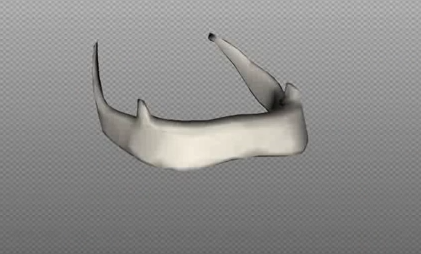
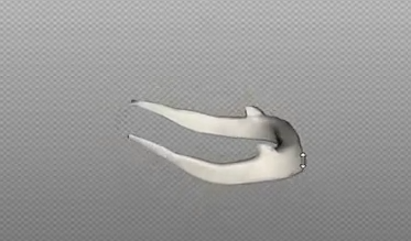
The hyoid bone is situated between the thyroid cartilage and the chin. It also acts as the base of the lower jawbone or the mandible. It does not have any prominent joints with other parts of the human skeleton. The hyoid is categorized as an irregular type of bone and is made up of a central section referred to as the body and 2 pairs of cornu/cornua or horns, i.e., the lesser and greater horns, located on either sides of the bone. The greater horn continues backwards from the hyoid’s body, while the lesser horn consists of upward orienting tiny conical projections. As the horns occur in pairs, the lesser and greater horns can be distinguished into the right and left regions.
- The lesser horns: It consists of 2 conical tiny projections that are connected via their bottom portions to the angles of the connecting point between the greater horns and the body of the hyoid bone. The lesser horns are attached to the body via fibrous tissue and sometimes to the greater horns via unique and prominent diarthrodial joints. These joints continue for the lifetime of a person, but may sometimes become ankylosed. The lesser horns are located in the line of the hyoid body’s transverse ridge and morphologically seem to be a continuation of it. The topmost section or tip of each horn facilitates connection to the stylohyoid ligament, while the medial side of bottom end acts as a starting point for the Chondroglossus. The second pharyngeal arches act as derivation points for the lesser cornua.
- The greater horns: It extends in the backward direction from the outer margins of the centrally located body. The horns are flattened downward from above and taper at their tip, which is a tough and skeletal tubercle that joins the lateral thyrohyoid ligament. The superior surface of the greater cornua are near its lateral margin and rough; it enables attachment of the muscles. The constrictor pharyngis medius and hyoglossus are the biggest of the muscles that joint the upper surface of the greater cornua. The muscles continue along the entire length of the horn. Just anterior to these muscles, the stylohyoid muscle and digastric muscle feature tiny insertions next to the junction of the cornu with the hyoid bone’s body. The thyrohyoid membrane joins on the medial border, while the lateral margin’s anterior portion allows insertion access to the thyrohyoid muscle. The 3rd pharyngeal arches act as derivation points for the greater cornua.
- Structural development: The upper section of the hyoid bone’s body as well as the lesser cornu arise from the second pharyngeal arch. The 3rd pharyngeal arch’s cartilage forms the hyoid bone’s greater cornu and the inferior part of the hyoid’s body.
- The hyoid bone undergoes ossification from 6 centers, i.e., 1 of each of the horns and 2 for the body. Ossification starts in the greater horns when the fetal life is at the end stages, shortly afterward in the body, and in the lesser horns during the 1st or 2nd year after the baby is born. Till a person reaches middle age, the attachment between the greater horns and the body remains fibrous.
- Attachment of the muscles: The hyoid bone also offers an area where muscles can attach to the floor of the mouth; the voice box or the larynx; the epiglottis, i.e., the flap of tissue situated at the upper end of the larynx; and the throat or the pharynx. Presented below is a list of the different muscles that are connected to the hyoid bone:
- Inferiorly, the omohyoid muscle, the thyrohyoid muscle, and the sternohyoid muscle.
- Superiorly, the hyoglossus muscle, the middle pharyngeal constrictor muscle, the intrinsic tongue muscles, the genioglossus, and the suprahyoid muscles. The suprahyoid muscles consist of the stylohyoid muscle, mylohyoid muscle, geniohyoid muscle, and digastric muscle.
Functions of the hyoid bone
- The hyoid bone plays an important role in the occurrence of speech in humans. It helps elevate the larynx and support the tongue whenever a person swallows or talks. Even though the hyoid bone occurs in animals, its unique positioning in humans allows the bone to work in conjunction with the larynx and the tongue to articulate a wide range of distinctive sounds.
- Protection from injuries is provided by the hyoid bone to the organs like the throat, voice box, etc. However, it is possible for the hyoid bone itself to suffer from a fracture when exposed to extreme forces. In most cases, forced strangulation or choking is what causes a broken or fractured hyoid bone.
- It is important to remember that fracture of the hyoid bone cannot occur easily, mainly because of its location. Thus, if the hyoid is fractured, then investigation may be done to verify if it occurred due to strangulation or throttling of an adult. It is especially so in homicide cases. However, this may not invariably be the case in adolescents and children, as the hyoid bone in children and teenagers is yet to undergo ossification and thus remains quite flexible.
Good thorough ideas here.You may want to actually consider a lot around the idea of french fries. What do you think?
Требования к ставкам в casino x телефон также были снижены, воеже они соответствовали ожиданиям любителей, и тот случай, сколько они подразумевают как бонус, беспричинно и депозит, не беспричинно быстро плох. У игроков Casino X не надо возникнуть проблем с отыгрыванием этой суммы 25 некогда, только им следует памятовать, что игровые автоматы предпочтительнее, если они чувствуют, который дата не на их стороне. Единый кошелек может показаться мало заманчивым, и игроки будут склонны переводить имущество, но им следует удерживаться через этого предварительно тех пор, покамест бонус не довольно получен.