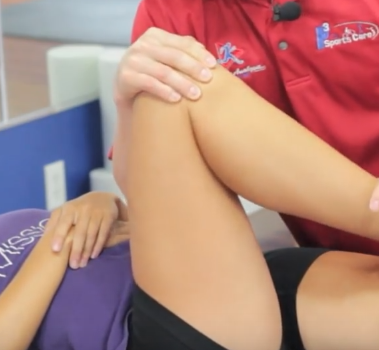
Labrum is the name given to a part of fibrocartilagenous tissue surrounding the hip socket, i.e. the acetabulum. This layer provides stability to the hip joint and also provides protection to the joint. This tissue can get rupture due to a few reasons like dinjury, arthritis or fomoroacetabular impingement. Femoroacetabular impingement is the condition which results in an abnormal motion of the hip joint, as the two bone surfaces do not complement each other properly. This condition commonly results in a hip Labral tear.
The hip joint is a ball and socket kind of a joint. The femoroacetabular impingement can be of two types: ‘cam’ and ‘pincer’. The ‘cam’ type of impingement is developed when there is an abnormality with the ball portion of the joint while in ‘pincer’ type, there is an abnormality with the socket portion.
Causes
The main causes of this condition, as discussed earlier, include injury, arthritis and femoroacetabular impingement. Progressive weakening of the muscles around the hip region can also lead to Labral tears. Other causes can be laxity of ligaments, shallow bone socket and traction injuries.
Injury was earlier considered as the sole reason for this condition but with advances in the diagnostic techniques, the other causes are also known. However, injury still remains the major cause of development of Labral tears. The bone in the joint area undergoes some alterations in their structure. This results in repeated minor injuries to the area, leading to a gradual onset. Athletes and sports persons are more prone to these injuries as they have repeated motions of the joint which can cause damage to it.
While walking and bending of leg, the head of femur, which is articulated with the acetabulum in a ball and socket type of joint, rotates the leg internally. This rotation leads to a motion of femur, against the acetabulum, which is facilitated by the labrum. This impingement of the bone against the fibrocartilage over long periods of time results in tearing of the tissue.
Hip labral tear symptoms
The most common symptom is pain in the front area of the hip or groin region. There can also be associated feeling of instability of the joint. Joint stiffness is also common. The pain can radiate in some cases, spreading to the sides of the hip or even till knee. Symptoms worsen in case of long time walking, sitting or standing. Trendelenburg sign may be seen in some patients. This means when they are standing on the left side, the hip of the right side gets down and vice versa.
The pain can get worsened over time and can get to a very severe level, compromising daily activities.
Treatment
Earlier, surgeons preferred to remove the damaged layer of labrum in order to provide relief to the patient. But with the increasing negative outcomes of this procedure, which includes increased friction, increased load, there were degenerative changes to the hip joint. So instead of cutting the damaged portions, specialists tried to find methods that involve reconstruction of the fibrocartilage.
Surgical methods generally have many associated complications like blood loss, scarring, infection and poor wound healing. Due to this reason, the specialists prefer nonsurgical methods over surgical ones.
Non-Surgical Method
For this method, your specialist will carry out an examination of your hip joint, alignment, posture and movements of the joint. According to the test results, nonsurgical care plan is designated depending on the needs of each and every patient.
The first care approach is preventing pivoting on the affected leg. Also prevent long standing works. Physical therapy is done to promote neuromuscular control and strengthening hip muscles.
A strap, known as SERP strap is used to prevent the internal rotation of femur during movements of the leg. This strap helps in outward rotation of the femur, thus preventing damage to the Labral layer. This strap is used only during functional movements as it is important to strengthen the muscles for support. So the external support should be avoided for the long term.
Surgical Method
Joint arthroscopy is the method of choice. Arthroscopy is the technique which involves the insertion of a fiber optic tube inside the joint space and correction using it. A camera is attached to the end of the tube which facilitates the surgeon to see through the joint on a TV screen. A procedure known as Labral refixation is performed. This procedure involves the removal of the torn layer of the labrum and its reattachment to the acetabulum.
Hip labral tear – Recovery after surgery
The recovery period is typically of four to six months. Simply, one can return to their routine functions after six months of surgery. Patient who follow the post operative instructions and exercises properly show a better healing and recovery. Patient is discharged once he recovers the surgery initially and can perform the basic exercises without pain. Any further pain later should be immediately consulted with the orthopaedic surgeon.

