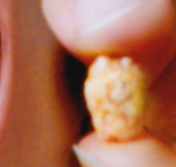
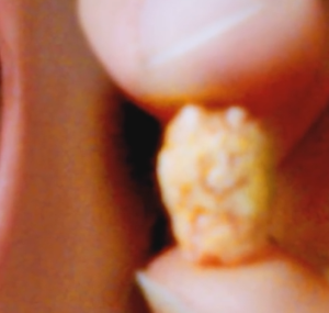
Saliva is a liquid which is constantly produced from the salivary glands of your body and flows to the oral cavity through ducts. Sometimes, some of the constituents of this saliva can get crystallized to form a solid mass and may get stuck in the gland body or the duct through which it flows. This results in swelling and pain in the neck.
These stones are usually less than 1 cm in diameter. However, they may be as small as less than 1 mm in diameter or as large as a few centimetres. Majority of the stone mass consists of calcium.
This stone usually needs to be removed by surgery. However, smaller stones can get removed by themselves or by slight probing.
Causes
There is not any exact known cause for the formation of these stones as the systemic calcium levels are not raised. Normal constituents of saliva include phosphates and calcium. These substances crystallise to form such stones. They obstruct the way of flow of saliva through salivary glands and thus cause swelling due to accumulation of saliva.
The exact reason for the formation of such stones is not known but certain factors can increase the risk. Such factors include:
- Dehydration as it causes your saliva to concentrate more and thus increasing the chances of crystallisation.
- Taking certain medications which reduce the salivary flow like anti hypertensive and antihistamines.
- Improper diet is also a cause of reduced salivary flow, as food induces salivary flow.
Location
There are three major paired salivary glands, namely the parotid, submandibular and sublingual. The salivary stones are most commonly seen in either of the submandibular glands or their ducts under the tongue. Formation of stones in the other two glands is comparatively rare.
Symptoms
In case of very small stones, you may not even be able to witness its presence as it will not show any symptoms. Larger stones may be visible when the patient opens his or her mouth. Such stones cause discomfort and show symptoms until it is removed.
You can witness varied types of symptoms depending on the size of the stone and degree of obstruction.
If the duct is partially obstructing the way of salivary flow, patient can present with symptoms like moderate swelling of the gland which will periodically subside and recur by themselves. There is associated dull pain in the affected gland which appears and subsides by itself. A salivary stone may also result in infection of the salivary gland which may further cause severe pain and discomfort.
If your salivary flow is completely obstructed from the gland, you may witness severe pain during mealtime. This pain is sudden and intense in nature. It gradually subsides over time and reappers during the next meal. Infection can also result in systemic complications like fever and malaise.
Swelling and pain are results of saliva make up in the gland which cannot escape due to obstruction of the duct.
Salivary Stone -Pictures
Diagnosis
It can be clinically seen during a routine oral examination. If it doesn’t show in that, radiographs might be helpful to diagnose its presence. If the stone does not get diagnosed in an X-ray, some further tests may be done to diagnose the condition:
- CT Scan, which shows details of the soft tissues and will show up any disorder in the same
- Sialography, which includes the insertion of a radiolucent dye into the substance of teh gland, which will show up on a radiograph taken after the insertion of the dye.
- Magnetic Resonance Imaging, also known as MRI, which uses a combination of magnetic fields and radio waves to produce image detailing.
- Sialendoscopy, which includes the insertion of a thin tube with a camera and light on its end. This tube is inserted into the salivary duct which enables the clinician to see through the duct. The stone can also be removed at the same time.
Removal of Salivary Stone
The clinician can make an attempt to remove the stone using a blunt thin instrument. Such an instrument is inserted into the canal and the stone can be removed thereby. If the stone does not get removed by this procedure, then a sialendoscopy is done. In this procedure, a sialendoscope is inserted in the duct of the gland after administration of local anaesthesia. The end of the tube has a small instrument for gripping and removing the stone. When the stone is observed, it is grabbed using this instrument and is pulled out of the canal. Only tiny stones can be removed using this procedure. In case of larger stones, they first need to be broken into smaller fragments using shock wave therapy, known as extracorporeal shock wave lithotripsy, and then the smaller pieces are removed one by one. Earlier, such stones were removed by making small incision in the oral cavity at the site of stone after administration of anaesthesia. But, this procedure is not required nowadays.


Накрутка Twitch Зрителей
Шоколадная плитка открытка Новосибирск